

If your vision becomes blurred again months or years after successful cataract surgery, the most common cause is "secondary cataract" — clouding of the capsule that holds the intraocular lens. It is not a cataract that has come back, and treating it is simple: a short YAG laser procedure in the clinic. Dr. Stern performs this treatment at Hadassah Ein Kerem Medical Center.
One of the most common misunderstandings among cataract patients concerns the concept of "secondary cataract."
The Medical Fact: You Cannot Develop Another Cataract After the Natural Lens Is Removed
Once the clouded natural lens has been surgically removed and replaced with an artificial intraocular lens, the original cataract cannot grow back. The artificial intraocular lens is a permanent implant and cannot develop the original cataract again.
However, some patients may develop gradual blurring months or years after an initially successful cataract surgery. This condition is medically called Posterior Capsule Opacification (PCO), and is sometimes referred to in everyday language as "secondary cataract" or "capsule opacification."
The Mechanism of PCO - The Sequence of the Process
- Residual lens epithelial cells (LECs) remain after surgery.
- The cells proliferate, migrate, and undergo fibrotic/myofibroblastic metaplasia.
- Elschnig pearls form - swollen, bladder-like Wedl cells - and/or fibrotic wrinkling occurs, causing light scatter and visual distortion.
- Ultimately, opacification of the central visual axis develops over the posterior capsule.
The Biological Mechanism of PCO
During cataract surgery, the surgeon carefully opens the front part of the natural lens capsule, but leaves the sides and back of the capsule - the capsular bag - intact, to support the new intraocular lens.
Despite thorough cleaning and polishing of the capsule during surgery, a small number of microscopic, viable lens epithelial cells (LECs) inevitably remain attached to the inner surface of the capsule. Over time, these cells can undergo the following processes:
- Proliferation and migration: The cells proliferate and migrate across the transparent posterior capsule, behind the optic of the artificial lens.
- Metaplasia: The epithelial cells can transform into myofibroblasts and begin depositing a matrix of fibrous tissue and collagen.
- Wrinkling and opacification: As the fibrotic collagen matrix contracts, microscopic wrinkles and physical opacification form on the posterior capsule. This distorts the path of incoming light and can cause symptoms similar to those of the original cataract.
Visual Patterns of PCO
- Elschnig pearls: A classic pattern in which LECs proliferate and form clusters of transparent, swollen, bladder-like cells, also called Wedl cells. They appear as tiny pearls scattered across the posterior capsule, scattering light and degrading image quality.
- Fibrotic metaplasia: A dense, white sheet or ring of fibrotic tissue, sometimes associated with contraction or phimosis of the anterior capsule, directly covering the central visual axis.
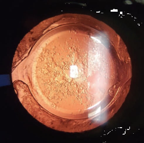
Incidence and Risk Factors
Thanks to modern IOL designs - especially hydrophobic acrylic materials and sharp, square posterior edges - the rate of PCO has dropped significantly.
- Older studies with round-edged lenses reported PCO rates of about 3% to 53% within three years of surgery.
- Modern clinical trials with hydrophobic, square-edged lenses report lower rates: up to 18.6% three years after surgery and up to 22.6% five years after surgery.
Risk factors for PCO include:
- Young age: In children and young people, the healing response is stronger and cell proliferation is more active, resulting in significantly higher PCO rates.
- History of uveitis or intraocular inflammation: Chronic inflammation promotes cell migration and metaplasia.
- Pseudoexfoliation syndrome: A systemic condition associated with fragile zonules and increased capsular instability.
- Incomplete cortex removal: Leaving significant cortical remnants after surgery provides a rich substrate for cell proliferation.
- Larger capsulorhexis: If the opening in the anterior capsule is larger than the IOL optic, the capsule cannot wrap snugly around the edges of the lens, impairing the physical barrier effect of the square edge.
- IOL material and round edges: PMMA or silicone lenses with round edges are associated with higher PCO rates compared with hydrophobic acrylic lenses with square edges.
Note: Studies cited in this guide indicate that no difference in PCO rates was found based on prolonged use of corticosteroid drops or nonsteroidal anti-inflammatory drops (NSAIDs) after surgery.
YAG Laser Posterior Capsulotomy - Treating Secondary Cataract
If posterior capsule opacification (PCO) develops, it can usually be treated quickly in the clinic with a non-invasive laser procedure called Nd:YAG laser posterior capsulotomy. The laser creates a precise central opening in the clouded posterior capsule to clear the visual axis, without contacting the intraocular lens. The success rate is very high, exceeding 95%, with immediate improvement in visual clarity.
When the Procedure Is Performed
YAG capsulotomy is appropriate when PCO impairs visual acuity or causes glare and halos that interfere with reading, driving, or work, and also when the opacity prevents proper monitoring of the retina and optic nerve. The procedure is very safe; in rare cases - such as active intraocular inflammation or unbalanced glaucoma - the condition must be stabilized before performing the laser.
How It's Performed
The procedure takes about five minutes, is painless, and is performed in the ophthalmologist's office - usually after drops to dilate the pupil and lower pressure, and sometimes using a stabilizing contact lens. After the procedure, patients wait at the clinic for about 30 to 60 minutes for an intraocular pressure check. Because of the dilating drops, vision in the treated eye may remain blurry for several hours, so driving is not advised that day; reading and other normal activities can usually resume once the dilation wears off. In the days following the procedure, several new floaters may appear in the field of vision - a benign, temporary phenomenon.
Possible Risks
YAG capsulotomy is very safe, but like any medical procedure, it carries rare risks: a temporary rise in intraocular pressure is the most common complication, and usually responds well to drops. Mild macular edema and minor damage to the intraocular lens are less common. The rarest but most significant complication is a retinal tear or detachment - so you should seek care promptly if you suddenly notice many new floaters, flashes of light, or a dark shadow in your field of vision.
Frequently asked questions
Can a cataract come back after surgery?
No. Once the natural lens has been removed, the original cataract cannot grow back. Blurring that appears months or years after surgery is usually due to posterior capsule opacification (PCO), which is treated with a laser.
Does YAG laser treatment hurt?
No. The treatment takes about five minutes, in the clinic and without pain, usually after drops to dilate the pupil and lower eye pressure.
Will I need the YAG treatment again?
Usually not. The laser creates a permanent opening in the capsule, and PCO should not return in the opened area.
When can I drive after a YAG treatment?
Not on the same day: the dilating drops blur vision in the treated eye for several hours. Reading and normal activities can resume once the dilation wears off.
Are floaters after YAG a danger sign?
A few new floaters in the days after treatment are a benign, passing phenomenon. But a sudden shower of floaters, flashes of light or a dark shadow in the field of vision require immediate medical attention.
How common is secondary cataract?
With modern lenses it has become much less common. Clinical trials with hydrophobic square-edged lenses reported rates of up to 18.6% at 3 years and up to 22.6% at 5 years after surgery.