

1. Understanding the Natural Lens and Cataract
To understand what cataract is, it's important first to understand how a healthy eye processes light.
The Anatomy of the Natural Lens
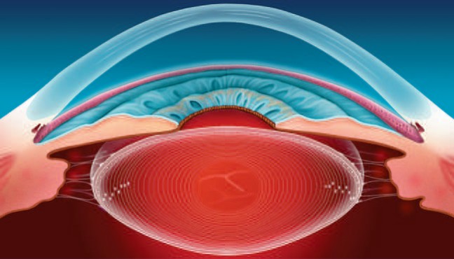
The human eye functions much like a high-quality camera. Light enters the eye through the transparent front window - the cornea - passes through the pupil, and is focused by a transparent, flexible structure called the natural, or crystalline, lens. The lens's main role is to focus light rays cleanly onto the retina - the light-sensitive tissue at the back of the eye, which functions like a camera's sensor. The retina converts light signals into electrical impulses and sends them to the brain, where they are interpreted as a clear image.
The natural lens is a transparent, biconvex structure located just behind the colored iris. It is held in place by a ring of delicate, thread-like fibers called the zonules of Zinn, which connect it to the surrounding ciliary body. The lens is made up of several distinct layers:
- Lens Capsule: An elastic, transparent, and extremely thin basement membrane that envelops the entire lens. The capsule protects the lens's internal structures while allowing nutrients to pass in and out.
- Lens Epithelium: A single layer of active cells located just beneath the anterior capsule. These cells are responsible for maintaining the lens's metabolic balance and continuously generating new lens fibers.
- Lens Cortex: The outer, younger layer of lens fibers surrounding the central nucleus.
- Lens Nucleus: The dense, older central core of the lens.
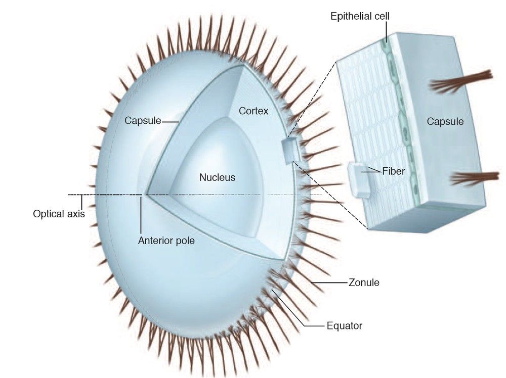
Unlike other organs, in which old cells are shed, the lens continues to produce new fiber cells at its periphery throughout life. The new fibers push the older fibers inward and compress them at the center of the lens.
What Is Cataract?
Cataract is not a film that grows over the surface of the eye, nor is it a tissue growth. Cataract is a gradual, progressive clouding of the natural lens itself.
In a young, healthy eye, the natural lens is completely transparent, allowing light to pass through it unobstructed. This transparency is maintained thanks to a highly organized arrangement of specialized proteins called crystallins. With age, or under the influence of various environmental and metabolic stress factors, these proteins undergo chemical and structural changes. They lose their organized arrangement, undergo denaturation - that is, a change or unfolding of their structure - and begin to clump together, forming water-insoluble aggregates.
These protein aggregates scatter light rays instead of allowing them to pass through cleanly. As the opacity becomes denser and spreads over a larger portion of the lens, it increasingly blocks and distorts the passage of light, and as a result vision becomes blurred, hazy, or dim.
Global Epidemiology: A Major Public Health Challenge
Cataract is a major health challenge worldwide and is considered a leading cause of blindness and visual impairment globally.
- According to the World Health Organization (WHO), more than 65.2 million people worldwide suffer from moderate to severe distance vision impairment or blindness due to untreated cataract.
- Data from the Global Burden of Disease study estimated that in 2015, approximately 12.6 million people were blind due to cataract, and that cataract accounted for a major share of the global burden of blindness. Up to 90% of these cases are concentrated in low- and middle-income countries, where access to eye care is limited.
- Approximately 20 million cataract surgeries are performed worldwide each year. Cataract surgery is one of the most commonly performed surgical procedures worldwide, and is considered one of the safest surgeries available today.
- Although most cases of cataract are age-related, cataract can also occur in infants. Congenital cataract occurs in about 1 in every 2,000 live births and accounts for about 5% to 20% of childhood blindness cases worldwide.
2. How Cataract Develops: Pathophysiology and Risk Factors
The development of cataract (cataractogenesis) is a slow, multifactorial biochemical process. Aging is the primary driver, but underlying cellular changes and various environmental factors can accelerate the process.
Why Cataract Develops With Age
With age, the structure of the natural lens gradually changes: the transparent materials that make it up lose their organized arrangement and begin to form cloudy areas, which scatter light instead of allowing it to pass through cleanly. As the opacity spreads over a larger portion of the lens, it increasingly blocks and distorts the passage of light, and vision becomes blurred.
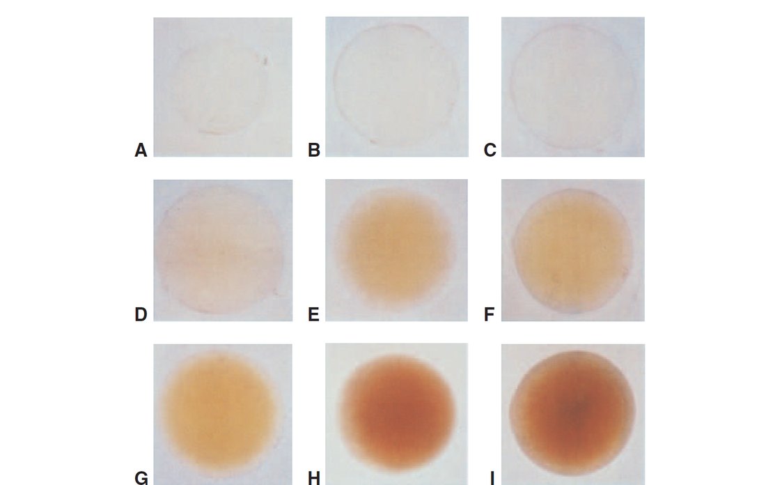
At the same time, the lens nucleus gradually changes color over the course of decades - from fully transparent to light yellow, and eventually to dark brown. This extreme color change is called brunescent cataract.
Systemic Disease: Diabetes as a Major Risk Factor
Certain systemic medical conditions can significantly accelerate the formation of cataract. The most prominent of these is diabetes mellitus.
In people with diabetes, cataract sometimes develops at a younger age and progresses faster than in people without diabetes - mainly because high blood sugar levels cause fluid to accumulate within the lens fibers, hastening the formation of opacity. In young people with poorly controlled diabetes, this process can sometimes cause acute diabetic cataract that progresses very rapidly.
Proven Medical and Environmental Risk Factors
Beyond aging and diabetes, several risk factors have been shown to be associated with an increased likelihood of developing cataract:
- Tobacco use: Smoking and smokeless tobacco use expose the body to toxins and cause significant oxidative damage to lens proteins.
- UV-B radiation exposure: Chronic, unprotected exposure to UV-B radiation from the sun promotes photo-oxidation and cross-linking of lens proteins.
- Systemic hypertension: High blood pressure has been statistically linked to an increased risk of cortical cataract.
- Long-term corticosteroid use: Whether taken systemically as pills, applied topically as eye drops, inhaled, or injected into the eye, corticosteroids are significantly associated with the rapid development of posterior subcapsular cataract.
- Eye trauma: Direct blunt trauma, such as a sports injury; penetrating injury; chemical burn; or exposure to ionizing radiation - all of these can cause cataract immediately or later on. Electrical injuries, such as a lightning strike or high-voltage workplace accidents, can also cause a characteristic type of cataract.
- Previous eye surgery: Patients who undergo vitrectomy - retinal surgery involving treatment of the vitreous - often develop rapidly progressing nuclear sclerotic cataract within months of the surgery.
- High myopia: Very high myopia is associated with earlier onset of nuclear cataract and posterior subcapsular cataract.
- Female sex: Epidemiological studies consistently show a slightly higher prevalence of visually significant cataract in women compared with men of the same age.
Diet and Dietary Supplements
Many patients ask whether cataract can be prevented or reversed through diet or dietary supplements. Studies in ophthalmology, including the Age-Related Eye Disease Study (AREDS) and AREDS2, have extensively examined various vitamins and minerals, such as lutein, zeaxanthin, vitamin C, and vitamin E.
A healthy diet rich in antioxidants is beneficial for overall eye health, but it has not been consistently proven that any medication or dietary supplement can prevent the development of cataract or slow its progression. Once a cataract has already formed, no eye drops, diet, or lifestyle change can dissolve the cloudy proteins and restore the lens's transparency. The effective and definitive treatment is surgical removal of the clouded lens.
3. Clinical Symptoms and Age-Related Types of Cataract
Cataract does not affect all patients the same way. Symptoms depend largely on the location of the opacity within the lens and how quickly it progresses.
Common Symptoms
- Decreased visual acuity: A gradual, progressive decline in the sharpness and clarity of vision, sometimes described as looking through a dirty window or through wax paper. This blurring cannot always be fully corrected with an updated glasses prescription.
- Glare and light sensitivity: Increased sensitivity to light. Bright sunlight can be very bothersome, and the headlights of oncoming cars at night can cause significant light scatter, making driving difficult and even dangerous.
- Reduced contrast sensitivity: A decreased ability to distinguish subtle differences between shades of gray or details in low-contrast conditions, for example when reading faded text or navigating a dim room.
- Refractive changes and "second sight": As the lens nucleus hardens and thickens, its refractive power increases, causing a shift toward myopia - that is, an increase in nearsightedness. In patients who previously needed reading glasses, including hyperopes or emmetropes, the temporary increase in near focusing power may suddenly allow reading without glasses. This temporary phenomenon is called "second sight." However, it is transient; as the cataract continues to cloud the lens, both near and distance vision gradually deteriorate.
- Monocular diplopia or polyopia: Double vision or the appearance of multiple ghost images in a single eye. This is caused by localized, irregular changes in the refractive index of different areas of the clouded lens, which split light rays before they reach the retina.
- Reduced color perception: As the lens yellows and darkens, it acts as a physical filter that blocks shorter wavelengths of light. As a result, color intensity decreases, particularly at the blue end of the visible spectrum, and colors may appear dull, faded, or yellowish. Because the change is usually gradual and affects both eyes, many patients are not aware of the decline in color perception until after cataract removal.
The Three Main Types of Age-Related Cataract
The three main forms of age-related cataract are classified according to their anatomical location within the lens. Many patients have components of more than one type at the same time.
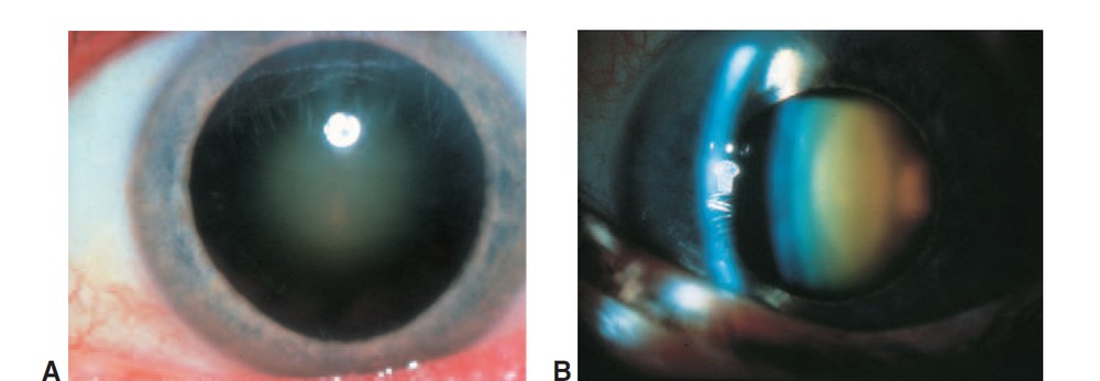
1. Nuclear Sclerotic Cataract
Anatomy: Gradual hardening (sclerosis) and progressive yellow-to-brown discoloration of the central lens nucleus.
Rate of progression: Very slow and gradual; it can take many years before the cataract becomes visually significant.
Main symptoms:
- Moderate to significant myopic shift - the temporary "second sight" phenomenon.
- Marked decrease in color perception, particularly in the blue-green range.
- Impaired visual function in low-light conditions - mesopic or scotopic conditions.
- Monocular diplopia, when the border between the hard nucleus and the soft cortex splits incoming light.
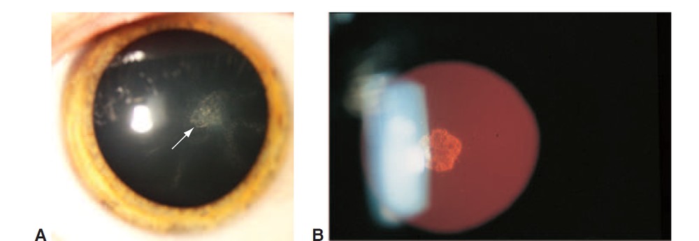
2. Cortical Cataract
Anatomy: The process begins as localized disturbances in the mature fiber cells of the lens cortex - the outer layer.
Pathophysiology: When the membrane integrity of the outer cells is compromised, essential metabolites are lost and protein oxidation, water absorption, and precipitation occur. The process typically begins at the periphery of the lens as fluid-filled spaces and "water clefts."
Clinical appearance: Over time, the opacities develop into wedge-like, or "spoke," shapes that progress from the edges of the lens toward the center.
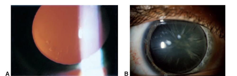
Main symptoms:
- Severe, limiting glare and light scatter, especially from strong point sources of light such as oncoming headlights at night.
- The effect on vision varies greatly depending on how close the "spokes" are to the central visual axis. If they remain at the periphery, vision can remain relatively clear; when they reach the center, the blurring becomes pronounced.
Stages of progression:
- Immature: The cortex is only partially clouded.
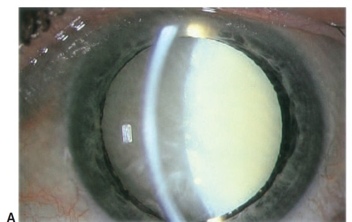
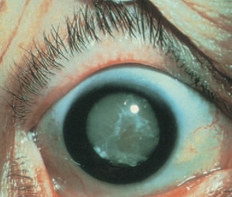
- Mature: The entire cortex, from the capsule to the nucleus, becomes completely white and opaque. The lens absorbs water, swells, and enlarges - a condition called intumescent cortical cataract. The swelling can crowd the eye's fluid drainage system and create a risk of angle-closure phacomorphic glaucoma - a medical emergency.
- Hypermature: Degenerated, liquefied cortical proteins begin to leak through an intact but wrinkled and shrunken capsule.
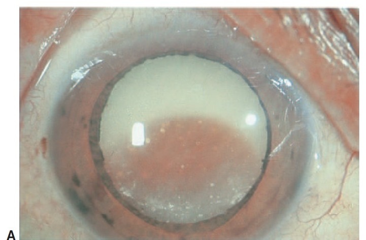
- Morgagnian: In an advanced stage of hypermaturity, the entire cortex liquefies completely and the dense, brown nucleus sinks freely to the bottom of the shrunken capsular bag.
3. Posterior Subcapsular Cataract (PSC)
Anatomy: Granular opacity that develops just in front of the posterior capsule of the lens, along the central visual axis.
Typical patients: Tends to occur in younger patients - sometimes in their 40s to 60s - compared with pure nuclear or cortical cataract.
Main causes: Strongly linked to corticosteroid use - topical, systemic, or inhaled - as well as eye trauma, intraocular inflammation (uveitis), exposure to ionizing radiation, high myopia, or heavy alcohol consumption.
Main symptoms:
- Pronounced, limiting glare in bright sunlight or brightly lit environments.
- Significant impairment of near vision and reading. During near tasks, such as reading, the pupil constricts, so light rays pass through the central, clouded area near the posterior capsule. As a result, near vision can be severely impaired, even when distance vision in dim lighting remains relatively normal.
Progression: The rate of progression varies greatly and can be much faster than that of nuclear or cortical cataract. A significant decline in vision can sometimes occur within just a few months.
4. Clinical Evaluation: What to Expect at Your Eye Examination
Before surgery can be planned, the ophthalmologist must perform a thorough, comprehensive eye examination to evaluate the cataract, determine whether surgery is indicated, and confirm that the rest of the eye is healthy.
1. Detailed Medical History
I begin with a conversation about your daily activities and how your vision affects your quality of life. We will discuss:
- Difficulty reading, watching television, or using a computer.
- Difficulty driving during the day due to bright sunlight, or driving at night due to glare from headlights.
- A history of falls or balance problems, since vision impairment from cataract has been statistically linked to an increased risk of falls and hip fractures in older adults.
- Systemic diseases such as diabetes; medications such as corticosteroids or prostate medications, for example tamsulosin, which can cause intraoperative floppy iris syndrome during surgery; and any previous eye trauma or eye surgery.
2. Comprehensive Eye Examination
- Visual acuity testing: Measuring best corrected visual acuity using a Snellen chart. If vision is blurry but can be corrected to 20/20 with new glasses, there is usually still no indication for surgery - unless significant, limiting glare is present.
- Slit-lamp biomicroscopy: Using a dedicated microscope to examine the structures of the front of the eye, including the cornea, iris, and natural lens. Using this examination, I assess the type and density of the cataract.
- Dilated fundus examination: Drops are used to dilate the pupil to allow a clear view of the back of the eye. A thorough examination of the macula, optic nerve, retina, and retinal blood vessels is essential. If advanced macular degeneration, a history of retinal detachment, or advanced glaucoma is present, the improvement in vision after cataract surgery may be limited. This examination helps me set realistic visual expectations.
3. High-Precision Preoperative Measurements - Biometry
Once a decision is made to proceed with surgery, the eye must be measured with great precision. This process is called biometry, and its purpose is to calculate the appropriate power of the intraocular lens (IOL) - by measuring the axial length of the eye and the curvature of the cornea, and using advanced calculation formulas that incorporate artificial intelligence to predict the most accurate outcome for each patient.
- Choosing a refractive target: We will discuss your visual goals. Is your goal to reduce dependence on glasses for distance vision as much as possible, or would you prefer very good near vision without glasses? This conversation guides the choice of lens power and the appropriate IOL type.
5. Indications for Surgery: When Is the Right Time?
Many patients are told they have "mild cataract" and ask when it is right to remove it. In modern ophthalmology, there is no single "right time," and there is no need to wait for the cataract to "ripen." The decision to operate is based mainly on two categories of indications: functional and medical.
1. Functional Indications - The Patient's Quality of Life
For most patients, cataract surgery is an elective procedure aimed at improving quality of life. The main indication is a situation in which the vision impairment caused by the cataract interferes with performing daily activities safely and comfortably.
Examples of functional indications:
- Inability to comfortably read books, newspapers, or digital screens, even with strong reading glasses.
- A feeling of insecurity driving at night due to glare and halos around headlights.
- Difficulty performing professional work, hobbies, or sports activities - such as golf or sewing - due to blurred or dim vision.
- Vision limits mobility, increases the risk of tripping or falling, or contributes to social isolation and depression.
Extensive clinical studies have shown that cataract removal significantly reduces the frequency of falls and hip fractures; observational cohort studies have also found an association between cataract surgery and lower mortality among older adults.
If the cataract is not bothering you and your vision is sufficient for your lifestyle needs, there is no need to have surgery right away. The cataract can be safely monitored, and you can proceed to surgery once it begins to affect your daily life.
2. Medical Indications
In certain situations, cataract surgery is necessary for medical reasons even if the patient is not bothered by their vision. These indications include:
- Phacolytic glaucoma: An inflammatory condition in which liquefied cortical proteins leak through the capsule of a hypermature cataract, clogging the eye's natural drainage system and causing a dangerous rise in intraocular pressure (IOP).
- Phacomorphic glaucoma: When an advanced intumescent cataract swells to the point of mechanically blocking fluid flow within the eye, causing acute angle-closure glaucoma - a severe, painful condition requiring urgent surgical intervention.
- Phacoantigenic uveitis: A severe, immune-mediated inflammatory reaction triggered by exposure of lens proteins following trauma or leakage from the capsule.
- Obscured view of the fundus: When the cataract is dense enough to prevent the ophthalmologist from viewing the back of the eye. A clear view of the retina is essential for diagnosing and monitoring significant eye diseases such as diabetic retinopathy, age-related macular degeneration, or glaucoma.
6. The Surgical Procedure: How Cataract Is Removed
Cataract surgery is one of the most commonly performed and successful surgical procedures in the world today, and one of the safest. More than 90% of patients achieve a postoperative result within 1.00 diopter of the planned refractive target.
Standard Phacoemulsification - The Modern Gold Standard
The primary surgical technique in high-income regions is phacoemulsification - a small-incision technique developed by Dr. Charles Kelman in 1967. In this method, ultrasound energy is used to break up - emulsify - the clouded lens and remove it through a small, self-sealing incision.
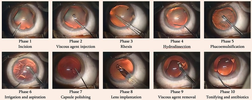
A brief overview of the phacoemulsification sequence:
- Anesthesia and pupil dilation.
- Creating a clear corneal incision.
- Capsulorhexis (CCC).
- Phacoemulsification of the lens nucleus.
- Irrigation and aspiration (I/A).
- Intraocular lens (IOL) implantation.
Step 1: Preparation and Anesthesia
The patient is brought into the operating room and the eye is prepared with disinfection. Local anesthetic drops are administered, and a small amount of intracameral anesthetic may also be given into the anterior chamber - the fluid-filled internal space between the cornea and the iris and lens - to ensure the surgery is comfortable and pain-free.
Step 2: Opening a Small Corneal Incision
I create a tiny incision, usually about 2-3 mm, at the edge of the clear cornea. The entire remainder of the surgery is performed through this incision, which self-seals at the end without the need for stitches.
Step 3: Opening the Lens Capsule
I gently open a round window in the thin capsule surrounding the natural lens, to allow access to the clouded nucleus inside.
Step 4: Breaking Up and Removing the Clouded Lens (Phacoemulsification)
Using an instrument that delivers gentle ultrasound energy, I break the clouded lens into small pieces and suction them out. I use the chopping technique, which breaks up the lens nucleus efficiently while using minimal ultrasound energy - which helps protect the eye's delicate tissues. After removing the central nucleus, I also gently remove the remaining softer outer cortex.
Step 5: Implanting the New Lens
A foldable intraocular lens is inserted through the same tiny incision and positioned within the capsular bag, where it unfolds and settles into place, where the natural lens was previously located.
Step 6: Completing the Surgery
I make sure the incision is well sealed, usually without the need for stitches, and administer antibiotic and anti-inflammatory drops to finish.
Femtosecond Laser-Assisted Cataract Surgery (FLACS)
In Femtosecond Laser-Assisted Cataract Surgery, or FLACS, an advanced, computer-guided laser is used to perform some of the initial steps of the surgery with great precision - opening the lens capsule and initial fragmentation of the nucleus - before phacoemulsification begins. Hadassah Medical Center has this type of advanced laser technology, and I recommend it to my patients: the laser adds precision to the key steps of the operation and reduces the ultrasound energy needed inside the eye.
In more detail: Laser cataract surgery (FLACS) — what’s the difference and who is it for
7. Postoperative Care: Medication Schedule and Recovery Guidelines
All the post-operative instructions are also gathered on a separate page: What not to do after cataract surgery — instructions and drop schedule
Vision is expected to gradually improve over the month following surgery. Postoperative eye drops are an important part of recovery and should be used exactly as prescribed, even if your vision already feels normal.
Which eye drops are used after cataract surgery? A typical tapering schedule
After surgery, three eye drops are typically prescribed, each gradually decreasing in frequency over about four weeks. Below is an example of a typical treatment schedule - always follow the specific instructions given to you by your surgeon, as the plan may vary depending on the characteristics of your surgery and eye:
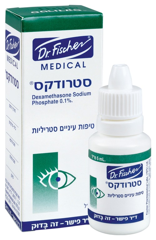
Dexamethasone Sodium Phosphate 0.1%
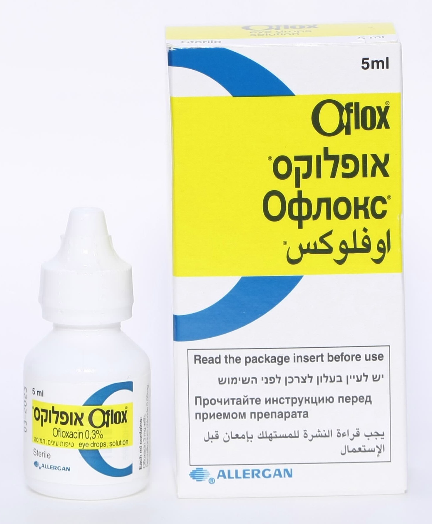
Ofloxacin 0.3%

Diclofenac Sodium 0.1%
| Drop | Day of surgery | Week 1 | Week 2 | Week 3 | Week 4 |
|---|---|---|---|---|---|
| Sterodex - anti-inflammatory steroid | Every hour | 6 times a day | 3 times a day | Twice a day | Once a day |
| Oflox - antibiotic | Every hour | 3 times a day | Stop | Stop | Stop |
| Dicloftil - anti-inflammatory (NSAID) | Every hour | 3 times a day | 3 times a day | 3 times a day | 3 times a day |
Pain after cataract surgery
You may take pain relievers as needed - acetaminophen, dipyrone, or ibuprofen (Nurofen) - provided you have no allergy or contraindication to these medications.
On the day of surgery
- It's recommended to rest at home; there is no restriction on movement.
- Do not shower on the day of surgery.
- Protect the eye with a shield until the following morning, and for two additional nights while sleeping.
- Bending over and lifting light loads is permitted.
What not to do after cataract surgery
- Avoid situations in which the eye could be struck, for example while playing with young children, pets or balls.
- Avoid sports involving a risk of trauma for at least one month.
- Shower with your head tilted back, to prevent water from entering the eye, for one week.
- Do not enter a pool, the sea, a mikveh, or standing water for one month.
Hygiene and eye care during recovery
- Do not touch or rub the eye.
- You may clean discharge using sterile eyelid pads, available at pharmacies.
- Wash your hands thoroughly before instilling any drop.
- Wait at least 5 minutes between different types of drops.
- Do not allow the bottle tip to touch the eye, eyelids or eyelashes when instilling the drops. If instilling drops is difficult, it's recommended to do so lying down.
8. Intraocular Lens Technologies and Implant Options
A comparison table and FAQs on choosing the lens: Types of lenses in cataract surgery
Cataract surgery is not just a procedure aimed at restoring transparency to the eye; it is also a personalized refractive procedure. When the clouded natural lens is removed, it must be replaced with a permanent artificial implant called an intraocular lens, or IOL.
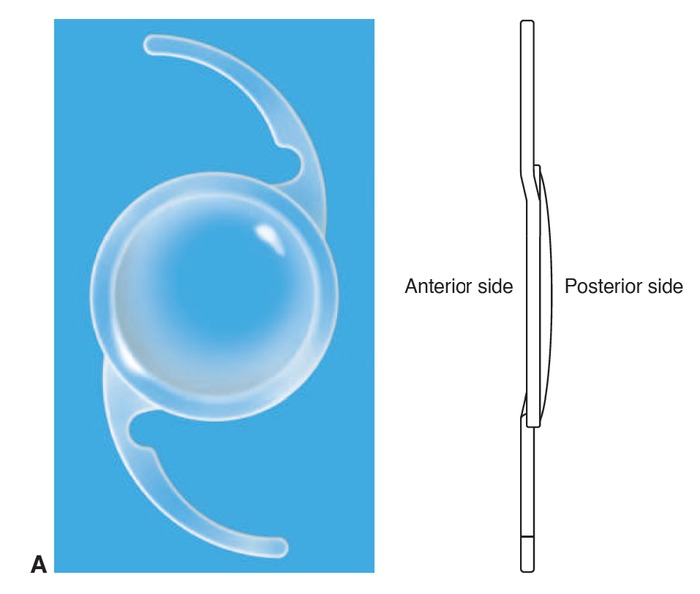
Modern IOL Materials and Design
Modern IOLs are made of soft, flexible materials - mainly acrylic, and sometimes silicone - that allow them to be folded and injected through a tiny incision. These materials are highly durable and designed to remain transparent within the eye for a lifetime. In patients who may need future retinal surgery involving silicone oil, I generally avoid implanting a silicone lens, since the oil can adhere to it and impair vision.
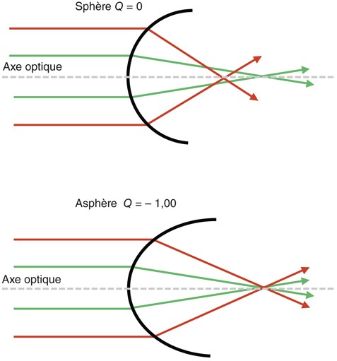
Lens design also has clinical significance: the sharp, square posterior edges common today significantly reduce the risk of later posterior capsule opacification (PCO) compared with older designs; and an "aspheric" optical design improves visual acuity and vision in low-light conditions, such as night driving.
Classification of Intraocular Lenses (IOL): Choosing a Focusing Range
Surgeons today have a wide range of IOL platforms available, allowing the visual outcome to be tailored to the patient's lifestyle, career, and personal hobbies.
Broadly speaking, there are three main groups:
- Monofocal lenses: A single, sharp focus point, usually for distance; reading glasses are needed in most cases.
- Astigmatism-correcting lenses: Toric lenses designed to correct existing astigmatism.
- Premium presbyopia-correcting lenses: Multifocal and EDOF options that provide additional ranges of vision.
1. Monofocal Lenses - The Traditional Option
Monofocal lenses are designed to provide very sharp, high-contrast vision at a single focal distance - usually set for distance vision, that is, emmetropia.
How it works: If both eyes are targeted for distance emmetropia, sharp, good vision can be expected for driving, watching television, and walking. However, since a monofocal lens cannot change shape and accommodate, reading glasses will usually be needed both for intermediate tasks - such as using a computer or cooking - and for near tasks, such as reading a book, using a phone, or sewing.
Targeting for myopia: Patients who have been nearsighted their whole lives and enjoyed reading without glasses can choose to target monofocal lenses for near vision - that is, for myopia. In this case, they will be able to read without glasses but will need glasses for distance vision, for example for driving and outdoor activities.
2. Toric Lenses - Astigmatism Correction
Astigmatism occurs when the cornea has different curvatures in different meridians, causing light to focus unevenly and producing blurred or distorted vision at all distances. About 40% of patients undergoing cataract surgery have corneal astigmatism of 1.00 diopter or more, and about 15% to 20% have significant astigmatism of 1.50 diopters or more - a level that usually justifies the use of a dedicated toric lens.
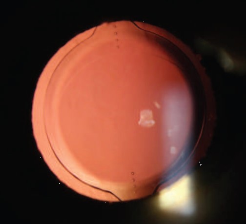
How they work: Standard IOLs do not correct astigmatism. A toric IOL is a specialized lens with different focusing powers in different directions. During surgery, I align the markings on the toric lens precisely with the patient's corneal astigmatism axis.
The importance of alignment: Precision is critical. For every one degree that a toric lens rotates away from its planned axis, it loses approximately 3% of its astigmatism-correcting power. I sometimes use advanced guidance systems during surgery to achieve the most accurate positioning possible.
For the disadvantages and limitations of toric lenses, see Question 7 in the FAQ.
Difficulty With Near Vision After Cataract Surgery, When the Target Is Distance Vision
Patients who choose a monofocal lens - standard or toric - targeted for distance vision may find that they need reading glasses after surgery. This is an expected result of the type of lens, not a complication. Some patients find over time that the dependence on reading glasses is more bothersome than they expected. The simplest and most common solution is to wear reading glasses; for those who wish to further reduce dependence on glasses after surgery, dedicated presbyopia-correction methods are available, detailed below.
Patients who are not satisfied with the refractive outcome after cataract surgery - for example, a larger-than-expected residual refractive error at any distance - should not have to settle for the result. There are two main approaches to correction: additional laser surgery on the cornea (PRK or LASIK) for fine-tuning the focus, or adding a supplementary intraocular lens (piggyback IOL) over the existing lens, without removing it. In select cases, surgical exchange of the intraocular lens itself may also be considered. The appropriate choice depends on the cause of the residual refractive error and the characteristics of the eye, and it's recommended to consult with your surgeon.
3. Methods for Correcting Presbyopia
Anyone who wants to reduce dependence on reading glasses - not just achieve astigmatism correction or sharp distance vision - can consider one of the following methods.
Monovision in Pseudophakia
Monovision is a personalized technique designed to reduce dependence on glasses using standard monofocal lenses.
How it works: I implant a lens targeted for distance in the dominant eye and a lens targeted for near vision in the non-dominant eye.
The result: The brain naturally combines the two images, so that one eye provides mainly distance vision and the other eye provides mainly near vision.
Variations: Traditional monovision creates a larger difference in focus, of about minus 1.50 to minus 2.50 diopters. This can provide very good near vision, but may slightly reduce depth perception. Mini-monovision, or modified monovision, uses a more moderate difference of about minus 0.50 to minus 1.50 diopters; this better preserves depth perception while still providing good intermediate vision, for example for computer use.
Multifocal Lenses - Bifocal and Trifocal Premium Lenses
Multifocal intraocular lenses (MFIOLs) are designed to provide clear vision at multiple distances simultaneously, thereby significantly reducing - and sometimes eliminating - dependence on glasses.
How they work: These lenses use diffractive or refractive concentric rings on the surface of the optic. The rings physically split incoming light: some light rays are focused for distance, and some for intermediate or near range. Modern trifocal lenses, such as the Alcon PanOptix, provide three distinct focal points: distance, intermediate vision (for example, computer distance), and near vision (reading distance).
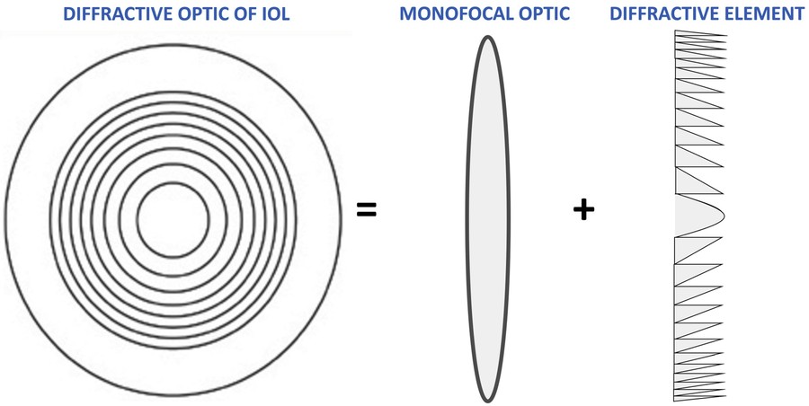
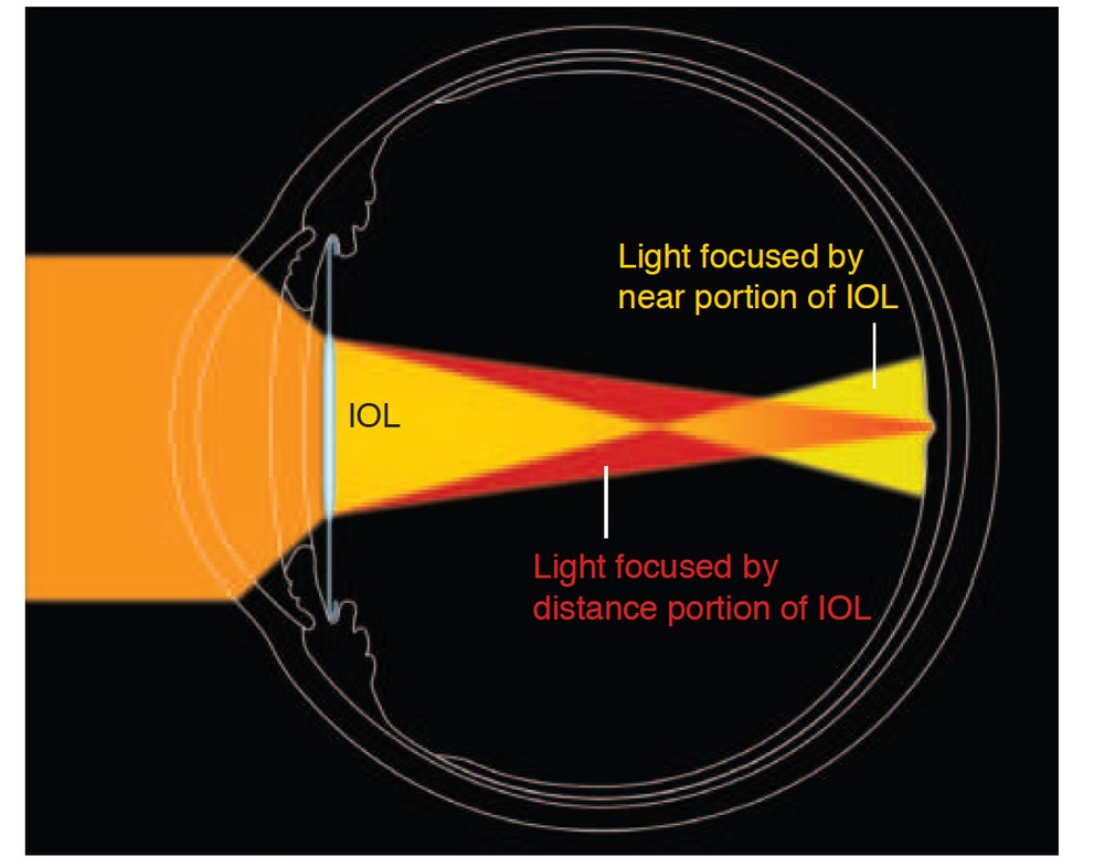
Drawbacks: Splitting light inherently causes some loss of contrast sensitivity, so images may appear slightly dimmer in dark rooms. In addition, the concentric rings can cause optical phenomena called dysphotopsias, including glare, halos, and starburst light scatter around light sources, especially when driving at night.
Patient selection: MFIOLs are premium lenses that require a very healthy eye. They are not recommended for patients with existing dry eye disease, corneal scarring, advanced glaucoma, macular degeneration, or epiretinal membranes, since these conditions can add to the impairment in contrast sensitivity and lead to an unsatisfactory visual outcome.
EDOF Lenses - Extended Depth of Focus
Extended Depth of Focus (EDOF) lenses are a modern advance in technologies designed to reduce dependence on glasses due to presbyopia.
How they work: Instead of splitting light into several separate, distinct focal points, as multifocal lenses do, EDOF lenses use dedicated diffractive or non-diffractive designs - such as Alcon's Vivity or Johnson & Johnson's Symfony - to stretch and extend the eye's single focal point, creating a continuous, extended range of vision.
Range of vision: EDOF lenses provide excellent, high-contrast vision at distance and intermediate range - for example, for the dashboard, computer, and countertop - with fewer halos and less glare compared with traditional multifocal lenses. With non-diffractive designs such as Vivity, the night-driving profile is largely similar to that of standard monofocal lenses; with diffractive designs such as Symfony, halos and glare are more common than with a monofocal lens, though less than with a trifocal.
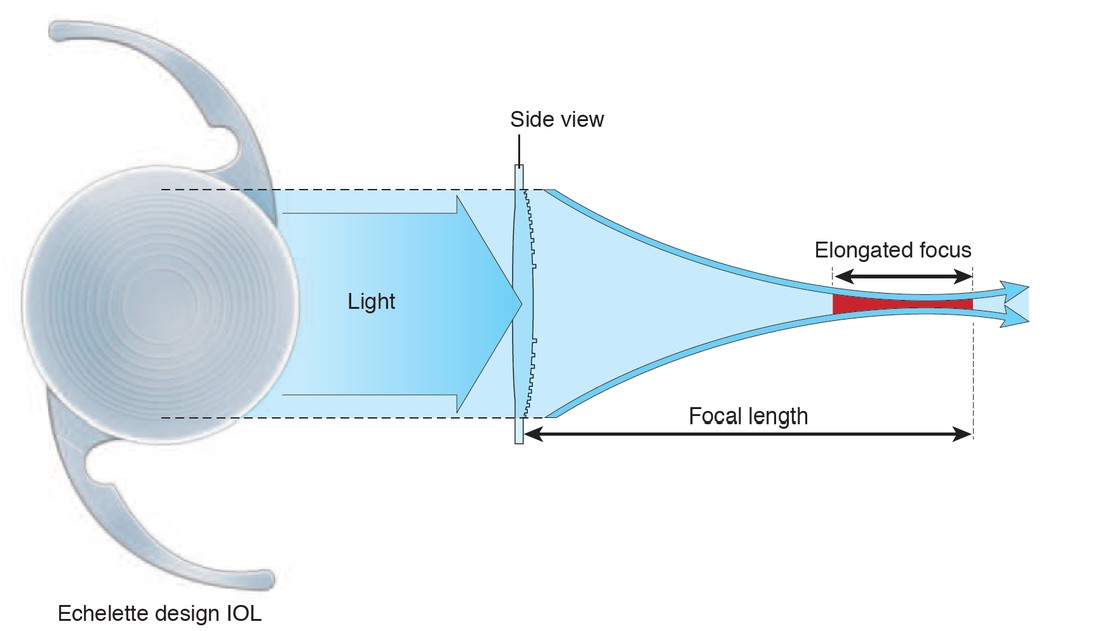
Near vision: Because the focus is elongated rather than split into a dedicated near point, EDOF lenses provide moderate near vision. Many patients can comfortably read large text but may need weak reading glasses for very small text or reading in dim light. To improve near vision, some surgeons combine mini-monovision with EDOF lenses, targeting the non-dominant eye for very mild myopia of about minus 0.50 diopters.
9. What Is "Secondary Cataract"? Posterior Capsule Opacification (PCO)
In brief, with FAQs about the treatment: Secondary cataract and YAG laser treatment
One of the most common misunderstandings among cataract patients concerns the concept of "secondary cataract."
The Medical Fact: You Cannot Develop Another Cataract After the Natural Lens Is Removed
Once the clouded natural lens has been surgically removed and replaced with an artificial intraocular lens, the original cataract cannot grow back. The artificial intraocular lens is a permanent implant and cannot develop the original cataract again.
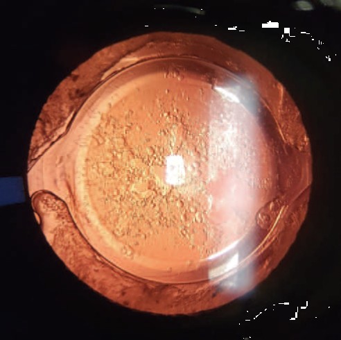
However, some patients may develop gradual blurring months or years after an initially successful cataract surgery. This condition is medically called Posterior Capsule Opacification (PCO), and is sometimes referred to in everyday language as "secondary cataract" or "capsule opacification."
The Mechanism of PCO - The Sequence of the Process
- Residual lens epithelial cells (LECs) remain after surgery.
- The cells proliferate, migrate, and undergo fibrotic/myofibroblastic metaplasia.
- Elschnig pearls form - swollen, bladder-like Wedl cells - and/or fibrotic wrinkling occurs, causing light scatter and visual distortion.
- Ultimately, opacification of the central visual axis develops over the posterior capsule.
The Biological Mechanism of PCO
During cataract surgery, the surgeon carefully opens the front part of the natural lens capsule, but leaves the sides and back of the capsule - the capsular bag - intact, to support the new intraocular lens.
Despite thorough cleaning and polishing of the capsule during surgery, a small number of microscopic, viable lens epithelial cells (LECs) inevitably remain attached to the inner surface of the capsule. Over time, these cells can undergo the following processes:
- Proliferation and migration: The cells proliferate and migrate across the transparent posterior capsule, behind the optic of the artificial lens.
- Metaplasia: The epithelial cells can transform into myofibroblasts and begin depositing a matrix of fibrous tissue and collagen.
- Wrinkling and opacification: As the fibrotic collagen matrix contracts, microscopic wrinkles and physical opacification form on the posterior capsule. This distorts the path of incoming light and can cause symptoms similar to those of the original cataract.
Visual Patterns of PCO
- Elschnig pearls: A classic pattern in which LECs proliferate and form clusters of transparent, swollen, bladder-like cells, also called Wedl cells. They appear as tiny pearls scattered across the posterior capsule, scattering light and degrading image quality.
- Fibrotic metaplasia: A dense, white sheet or ring of fibrotic tissue, sometimes associated with contraction or phimosis of the anterior capsule, directly covering the central visual axis.
Incidence and Risk Factors
Thanks to modern IOL designs - especially hydrophobic acrylic materials and sharp, square posterior edges - the rate of PCO has dropped significantly.
- Older studies with round-edged lenses reported PCO rates of about 3% to 53% within three years of surgery.
- Modern clinical trials with hydrophobic, square-edged lenses report lower rates: up to 18.6% three years after surgery and up to 22.6% five years after surgery.
Risk factors for PCO include:
- Young age: In children and young people, the healing response is stronger and cell proliferation is more active, resulting in significantly higher PCO rates.
- History of uveitis or intraocular inflammation: Chronic inflammation promotes cell migration and metaplasia.
- Pseudoexfoliation syndrome: A systemic condition associated with fragile zonules and increased capsular instability.
- Incomplete cortex removal: Leaving significant cortical remnants after surgery provides a rich substrate for cell proliferation.
- Larger capsulorhexis: If the opening in the anterior capsule is larger than the IOL optic, the capsule cannot wrap snugly around the edges of the lens, impairing the physical barrier effect of the square edge.
- IOL material and round edges: PMMA or silicone lenses with round edges are associated with higher PCO rates compared with hydrophobic acrylic lenses with square edges.
Note: Studies cited in this guide indicate that no difference in PCO rates was found based on prolonged use of corticosteroid drops or nonsteroidal anti-inflammatory drops (NSAIDs) after surgery.
10. YAG Laser Posterior Capsulotomy - Treating Secondary Cataract
If posterior capsule opacification (PCO) develops, it can usually be treated quickly in the clinic with a non-invasive laser procedure called Nd:YAG laser posterior capsulotomy. The laser creates a precise central opening in the clouded posterior capsule to clear the visual axis, without contacting the intraocular lens. The success rate is very high, exceeding 95%, with immediate improvement in visual clarity.
When the Procedure Is Performed
YAG capsulotomy is appropriate when PCO impairs visual acuity or causes glare and halos that interfere with reading, driving, or work, and also when the opacity prevents proper monitoring of the retina and optic nerve. The procedure is very safe; in rare cases - such as active intraocular inflammation or unbalanced glaucoma - the condition must be stabilized before performing the laser.
How It's Performed
The procedure takes about five minutes, is painless, and is performed in the ophthalmologist's office - usually after drops to dilate the pupil and lower pressure, and sometimes using a stabilizing contact lens. After the procedure, patients wait at the clinic for about 30 to 60 minutes for an intraocular pressure check. Because of the dilating drops, vision in the treated eye may remain blurry for several hours, so driving is not advised that day; reading and other normal activities can usually resume once the dilation wears off. In the days following the procedure, several new floaters may appear in the field of vision - a benign, temporary phenomenon.
Possible Risks
YAG capsulotomy is very safe, but like any medical procedure, it carries rare risks: a temporary rise in intraocular pressure is the most common complication, and usually responds well to drops. Mild macular edema and minor damage to the intraocular lens are less common. The rarest but most significant complication is a retinal tear or detachment - so you should seek care promptly if you suddenly notice many new floaters, flashes of light, or a dark shadow in your field of vision.
11. Frequently Asked Questions for Cataract Patients
Question 1: Does cataract surgery hurt?
No. Cataract surgery is performed under local topical anesthesia, using numbing drops and gel, and is sometimes combined with light sedation to help you relax. You may feel a mild sensation of cold fluid or gentle pressure on the eye, but there should be no pain. Many patients are surprised by how quick and comfortable the procedure is.
Question 2: Can cataract come back after surgery?
No. Once the natural lens has been removed, the original cataract cannot grow back. The new artificial lens - the IOL - is not the natural lens that clouded over. If vision becomes blurry months or years after surgery, the common cause is posterior capsule opacification (PCO) - clouding of the membrane behind the lens - a common, treatable condition.
Question 3: What is "second sight"?
"Second sight" is a temporary improvement in near vision that can occur during the development of nuclear cataract. Hardening of the lens nucleus increases its refractive power, making the eye more nearsighted, so people who previously needed reading glasses can suddenly read without them. As the cataract and the opacity continue to progress, both near and distance vision eventually become blurry.
Question 4: How long does recovery from cataract surgery take?
Most patients notice significant improvement in vision within 24 to 48 hours of surgery, although during the initial recovery period it's normal to experience mild grittiness, redness, and blurring. The eye heals fully and vision usually stabilizes within about 3 to 4 weeks, at which point new glasses can be fitted if needed.
Question 5: Can cataracts be prevented with vitamins or dietary changes?
It has not been proven that any vitamin, dietary supplement, or eye drop can prevent cataract formation or slow its progression once it has begun. A diet rich in antioxidants, such as leafy greens and colorful vegetables, is good for overall eye health, but the extensive scientific studies mentioned in this guide do not support the ability to prevent cataract through supplements or drops.
Question 6: Can cataract surgery be performed on both eyes at the same time?
Surgeons generally prefer to operate on one eye at a time, spacing the surgeries about one to two weeks apart. This ensures that the first eye has healed and has functional vision before operating on the second eye, while also minimizing the very rare risk of bilateral complications.
Question 7: What are the disadvantages of a toric lens?
A toric lens is a very effective way to correct astigmatism, but like any option it has limitations worth knowing in advance:
- Cost: A toric lens involves an additional charge compared with a standard monofocal lens.
- Lens rotation: In the first days and weeks, the lens may rotate slightly away from the planned axis, losing part of its correction (about 3% per degree). In a small minority of cases, a short procedure is needed to rotate the lens back into position.
- Residual astigmatism: The result depends on the accuracy of the preoperative measurements, and a small amount of astigmatism sometimes remains, which can be corrected with glasses or a laser enhancement.
- It does not correct near vision: A monofocal toric lens is focused at one distance, so reading glasses are usually still needed — unless a multifocal or EDOF toric lens is chosen.
- Not suitable for every eye: With irregular astigmatism — for example in advanced keratoconus or after corneal scarring — a toric lens may not be the right choice.
For most patients with significant astigmatism, the benefit — sharper vision and less dependence on glasses — clearly outweighs these limitations. The decision is made together with the surgeon, based on the preoperative measurements.
Question 8: What should you avoid after cataract surgery?
In the first weeks it is important to protect the eye: do not touch or rub it; wear a protective shield on the first night and the two nights after; do not shower on the day of surgery, and for the following week shower with your head tilted back so that water does not get into the eye; do not enter a pool, the sea, a mikveh or standing water for a month; and avoid sports and activities in which the eye could be injured for at least a month. Bending and lifting light loads are allowed. Most important of all, continue the drops exactly as scheduled, even when your vision already feels good. These are general instructions; always follow those you received from your surgeon. Full instructions and drop schedule →
The Key Difference: Cataract vs. "Secondary Cataract"
In summary, it's important to distinguish between these two conditions for patients preparing for surgery or recovering from it:
- Primary cataract: Clouding of the eye's natural lens. Treatment is surgical removal of the lens and replacement with an artificial implant - an IOL. Once the natural lens has been removed, that same procedure to remove the natural lens is not performed again in that eye.
- "Secondary cataract" - PCO: Opacification of the transparent membrane - the capsular bag - that holds the artificial lens in place, caused by migration of microscopic cells. Treatment is performed in the office, in under five minutes, using a non-invasive Nd:YAG laser. The laser creates a permanent opening in the membrane, and after treatment, PCO should not recur in that same opened area.
If you experience any change in your vision, it's recommended to schedule a comprehensive eye exam with an ophthalmologist. Today's advanced cataract surgery and premium lens technologies offer a significant opportunity for visual rehabilitation, and sometimes for achieving very good visual quality after surgery.