

1. Understanding the Eyes: An Introduction to Pterygium
It is entirely natural to feel concerned when you notice a new growth or a change in the appearance of your eyes. Many patients worry that a small, wing-shaped membrane appearing on the white of the eye could be a potentially serious condition. In medical terms this condition is called a pterygium. A pterygium is a wing-shaped fibrovascular growth that originates in the conjunctiva — the transparent membrane covering the white of the eye — and advances onto the cornea, the eye's transparent "window." Although it is indeed a growth of tissue, it is important to distinguish medical facts from common concerns.
In everyday language, patients describe a pterygium in many ways — "a film over the eye," "a growth on the white of the eye," or "a fleshy growth on the eye" — and it is sometimes called "surfer's eye" because of its link to sun and wind exposure. The name itself comes from the Greek pterygion, meaning "little wing," after its shape.
Myths vs. Clinical Facts
| Myth | The Clinical Fact |
|---|---|
| It is a form of eye cancer. | It is a benign, noncancerous lesion that generally grows slowly. |
| It will eventually cause total blindness. | It can affect vision by inducing astigmatism or blocking the visual axis, but in most cases it can be treated effectively. |
| It is "skin" growing over the eye's surface. | It is a degenerative process of the eye's own conjunctival tissue, often linked to environmental stress. |
| Once removed, it never comes back. | There is a risk of recurrence, which is why modern surgical techniques place strong emphasis on preventing it from returning. |
Understanding the basic anatomy of the eye's surface is the first step toward understanding the treatment options available.
2. What Exactly Is a Pterygium? Anatomy and Science
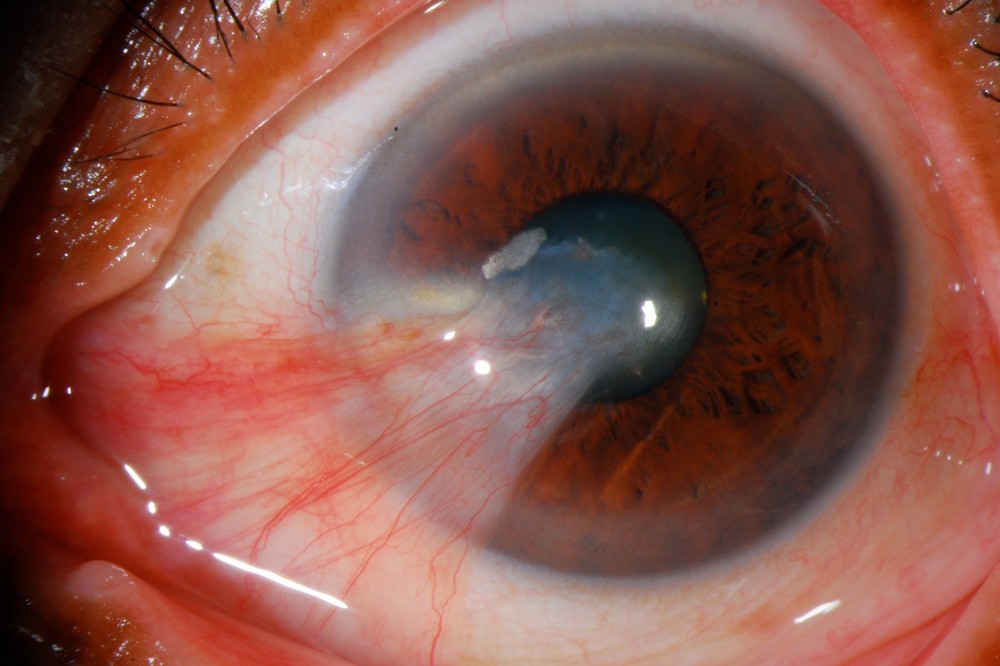
To understand pterygium, it helps to know the delicate structures of the eye's surface. The eye is protected by the conjunctiva, a thin membrane that lines the eyelids and covers the sclera — the white part of the eye. Where the sclera meets the transparent cornea lies an important transition zone called the limbus. This zone contains the Palisades of Vogt — radial ridges that contain important limbal stem cells that help renew the corneal epithelium. The cornea itself is a complex structure made of five layers, like a transparent, multi-layered pane that gives it extraordinary strength and clarity.
The Five Layers of the Cornea
- Epithelium: the outermost layer, serving as a protective barrier.
- Bowman's layer: a strong, acellular layer made of collagen fibers.
- Stroma: the middle and thickest layer, making up about 90% of the cornea's thickness and providing it with structural stability.
- Descemet's membrane: a thin, strong inner basement membrane.
- Endothelium: the innermost layer, functioning as a pump that maintains corneal clarity by regulating fluid.
The pterygium disrupts these layers — particularly the epithelium and Bowman's layer — as it advances from the conjunctiva toward the center of the cornea.
A Guide to Clinical Terms
- Stocker Line: a curved, vertical line of iron deposition in the corneal epithelium, sometimes seen just ahead of the head of a stable pterygium, used as a marker of stability.
- Pseudopterygium: unlike a true pterygium, this is a bridge of tissue that adheres to the cornea following trauma or inflammation. The two conditions can be distinguished with a clinical probe test: in a pseudopterygium, a small probe can be passed underneath the tissue at the limbus, whereas a true pterygium is attached to the tissue beneath it.
- OSSN Screening: to rule out Ocular Surface Squamous Neoplasia (OSSN), AS-OCT (Anterior Segment Optical Coherence Tomography) is used. Clinicians look specifically for hyperreflective epithelial thickening, which helps distinguish a benign growth from a lesion that may require further evaluation for ocular-surface neoplasia.
Pinguecula vs. Pterygium
The two conditions are often confused, but they behave differently:
| Feature | Pinguecula | Pterygium |
|---|---|---|
| Location | Confined to the conjunctiva — the white of the eye. | Advances onto the transparent cornea. |
| Shape | A yellowish, raised lesion. | A wing-shaped fibrovascular tissue. |
| Effect on vision | Minimal to none. | Can cause significant astigmatism or reduced vision. |
Grades and Classification of Pterygium
An ophthalmologist describes a pterygium by two main features: how far it has advanced onto the cornea, and how thick and active the tissue is. This classification helps decide between observation and surgery, and helps estimate the risk of recurrence after surgery.
By extent of corneal involvement
| Grade | Position of the pterygium's head | Clinical significance |
|---|---|---|
| Grade 1 | Between the limbus and a point midway between the limbus and the pupil margin. | Usually observation and conservative treatment, unless symptoms are significant. |
| Grade 2 | Between that midway point and the pupil margin. | Close to the visual axis and may cause astigmatism; surgery is considered depending on progression and symptoms. |
| Grade 3 | Crosses the pupil margin. | Affects the visual axis; surgery is recommended in most cases. |
By appearance of the tissue (Tan classification)
| Type | Appearance | Significance |
|---|---|---|
| T1 – Atrophic | Thin tissue; the episcleral blood vessels underneath are clearly visible. | Less active; relatively low risk of recurrence. |
| T2 – Intermediate | The underlying vessels are partly visible. | Moderate activity. |
| T3 – Fleshy | Thick, fleshy tissue that completely hides the vessels underneath. | More active and more likely to recur after surgery. |
A distinction is also made between a primary pterygium and a recurrent pterygium — one that has grown back after previous surgery. A recurrent pterygium tends to be more active and requires careful surgical planning. In the International Classification of Diseases (ICD-10), pterygium is coded H11.0.
3. What Causes Pterygium? Pathophysiology and Risk Factors
The development of a pterygium is linked to a process called elastotic degeneration, in which collagen in the eye's tissues breaks down and is replaced by abnormal fibers. An important clinical observation is that pterygium is significantly more common on the nasal side — the side of the eye closer to the nose — than on the temporal side. The reason for this asymmetry is not fully understood. One proposed mechanism is peripheral light focusing, in which oblique ultraviolet radiation passing through the temporal cornea may become concentrated near the nasal limbus. However, this mechanism may not fully explain the strong nasal predominance, and other factors have also been proposed.
The Four Main Environmental Risk Factors
- UV radiation exposure: the primary driver of cellular damage.
- Dry climate: lack of moisture places stress on the eye's surface.
- Dust: causes chronic irritation from airborne particles.
- Wind: increases tear evaporation and inflammation of the eye's surface.
This combination of factors is especially common in the so-called "Pterygium Belt" — a geographic zone near the equator characterized by high UV radiation. These environmental factors contribute to ocular-surface irritation, inflammation and the development or progression of pterygium.
4. Symptoms: How Pterygium Affects the Eye and Vision
As the pterygium grows, it disrupts the tear film — the fluid layer made of mucus, an aqueous component and lipids that keeps the eye's surface lubricated. As a result, patients may experience:
- Foreign body sensation: a feeling as if there is sand in the eye.
- Burning and redness: caused by local inflammation.
- With-the-rule or irregular astigmatism: as the pterygium exerts traction on and distorts the cornea, it flattens the horizontal meridian and can induce with-the-rule or irregular astigmatism, leading to blurred vision that may not be fully corrected with standard glasses.
The Connection to Cataracts
If both pterygium and cataract are present, timing matters greatly. Pterygium surgery should be performed first. After removal, I generally allow approximately three months for corneal measurements to stabilize before final cataract biometry, to ensure the most accurate result and the best possible visual outcome.
5. Non-Surgical Treatment and Prevention
For many patients, treatment begins with conservative measures aimed at reducing irritation and slowing progression.
Medical Treatment Protocol
- Lubrication: preservative-free artificial tears.
- UV protection: wraparound sunglasses with 100% UVA/UVB protection, and wide-brimmed hats.
- Short-term steroids: mild steroid drops to calm significant inflammation.
6. When Is It Time for Surgery? Surgical Indications
Surgery may be considered in the following situations:
- Threat to the visual axis: the growth is approaching the center of the pupil.
- Induced astigmatism: significant with-the-rule or irregular astigmatism causing blurred vision.
- Chronic discomfort: persistent redness and irritation that does not respond to drops.
- Restricted eye movement: the growth prevents the eye from moving freely.
- Cosmetic concern: the appearance causes the patient significant distress.
7. Understanding the Surgery: Modern Techniques Step by Step
Modern surgery is performed as an outpatient procedure under local anesthesia. The patient is awake during the surgery, but the eye is completely numbed.
Comparison of Surgical Techniques
| Technique | Recurrence Rate | Clinical Note |
|---|---|---|
| Bare Sclera | 32%–88% | High failure rate; the white of the eye is left "bare." |
| Amniotic Membrane Transplant (AMT) | 7%–69% | Uses processed tissue; success rates vary. |
| Conjunctival Autograft (CAG) | 1%–17% | Uses the patient's own healthy tissue; considered the gold standard for primary pterygium. |
| Conjunctival-Limbal Autograft (CLAU) | 0%–7% | Includes limbal stem cells; mainly reserved for recurrent or large pterygia. |
The exposed area is covered with healthy conjunctival tissue: either a conjunctival autograft (CAG), or a conjunctival flap, in which adjacent conjunctiva is slid over to cover the area without harvesting a graft from elsewhere on the eye, usually combined with mitomycin C. The tissue is secured with fine sutures; some centers use fibrin glue instead.
8. Surgical Adjuncts — Mitomycin C
During surgery, Mitomycin C (MMC) is often used. It is a potent antimetabolite — a medication that prevents cells, particularly fibroblasts, from dividing and forming scar tissue. In the past, using MMC in drop form carried risks; the modern approach is a brief, controlled intraoperative application directly to the treatment area, aimed at preventing the growth from returning.
9. The Road to Recovery — What to Expect After Surgery
- The first three days: a bandage contact lens (BCL) is fitted to protect the eye's surface during healing.
- From the first to the fourth week: vision gradually stabilizes as the eye heals.
- From the first to the third month: this is an important stage of gradual steroid tapering, over a period of four to twelve weeks. This is not merely for comfort; it is essential to preventing rebound inflammation, which is one of the leading causes of pterygium recurrence.
10. Risks and Safety Information
| Possible Risk | Prevention / Management |
|---|---|
| Recurrence | Covering the area with conjunctival tissue (autograft or conjunctival flap) and using mitomycin C. |
| Dellen (dryness-related thinning) | Managed with lubrication and a bandage contact lens (BCL). |
| Pyogenic granuloma | A small growth of scar tissue. Treated with topical steroids. |
| Muscle injury | Rare; avoided through careful identification and preservation of the relevant anatomical structures during surgery. |
11. Frequently Asked Questions for Patients
Is the surgery painful?
The procedure itself is not painful. For several days after surgery you may feel a "scratchy" or gritty sensation, which is managed with drops and the therapeutic contact lens.
When can I return to work or exercise?
Most patients return to light work within a few days. Strenuous exercise, contact sports and swimming should be avoided for at least two weeks.
When can I start using makeup again?
You should wait at least two to three weeks, until the surgical area has healed well, to avoid irritation.
How can I protect the results of the surgery?
Consistent protection from UV radiation with sunglasses, together with lubrication of the eye's surface, are the best long-term protections.
Can a pterygium go away with eye drops?
No. Lubricating drops — and, when needed, a short course of anti-inflammatory drops — relieve redness and irritation, but they do not dissolve the tissue. The only way to remove a pterygium is surgery. That said, a small, quiet pterygium usually does not need surgery: monitoring, UV protection and drops as needed are generally enough.
What are the common side effects after pterygium surgery?
In the first days and weeks, redness at the surgical site, a foreign-body or "scratchy" sensation, tearing and light sensitivity are common, sometimes with mild blurring. These are expected, temporary parts of healing and improve gradually; the redness usually fades within a few weeks. More significant complications are rare and are described in the Risks and Safety Information section.
Does a pterygium come back after surgery?
There is a risk of recurrence, and it depends mainly on the surgical technique. After simple excision alone (the bare sclera technique), recurrence rates are high. That is why the area is now covered with healthy conjunctival tissue — an autograft or a conjunctival flap — usually combined with mitomycin C, which lowers the risk substantially, to just a few percent in many series (see the comparison table in section 7). Following the gradual tapering of drops after surgery and protecting the eye from UV radiation reduce the risk further.
Summary
From initial diagnosis to advanced reconstructive techniques, modern pterygium treatment is highly successful. By understanding the anatomy of the eye and following a precise postoperative plan — including the gradual and essential tapering of steroids — patients can expect a healthy ocular surface and stable vision. Modern pterygium surgery aims not only to remove the lesion but also to restore a stable, healthy ocular surface while minimizing the risk of recurrence.