

1. What is an ICL lens?
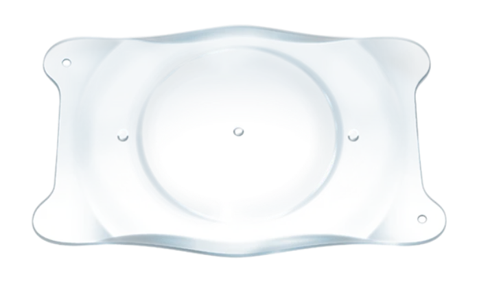
The ICL is a tiny, soft intraocular lens made from a biocompatible proprietary material called Collamer, a combination of collagen and polymer materials. Unlike the intraocular lens implanted during cataract surgery, the ICL does not replace the eye's natural lens. It is implanted inside the eye while the natural crystalline lens remains in place, fully preserving its natural focusing ability (accommodation) - which is why this procedure is called "phakic," meaning lens-preserving.
The lens is designed mainly to correct moderate-to-high myopia (nearsightedness), and in its toric version, accompanying corneal astigmatism as well. The modern model in use today, the EVO Visian ICL, includes a tiny central port 360 microns in diameter that allows intraocular fluid to flow through it naturally - an innovation that has made the surgery safer and simpler compared to older models.
2. How the lens works inside the eye
The ICL is implanted in the sulcus - the narrow space between the iris (the colored part of the eye) and the natural lens - not within the cornea and not in place of the natural lens. The ICL is positioned behind the iris and in front of the natural lens, with a small space - known as the vault - between the ICL and the crystalline lens, so the new lens does not touch the natural lens. The recommended postoperative vault ranges from about 250 to 750 microns. Preoperative measurements are used to select the appropriate ICL size and estimate the expected postoperative vault; after implantation, the actual vault can be measured using slit-lamp examination or anterior-segment imaging such as OCT or ultrasound biomicroscopy (UBM). This measurement is important because too low a vault increases the risk of developing a cataract, while too high a vault can cause a rise in intraocular pressure.
Because the natural lens remains in place, the eye continues to focus naturally at near and far distances in younger patients, exactly as it did before surgery.
3. Who it's suitable for: indications and patient selection
The ICL is mainly suitable for two groups of patients:
- Myopia too high for safe laser correction: The EVO Visian ICL is FDA-approved for the full correction of myopia from -3.00 to -15.00 diopters (and partial reduction up to -20.00 diopters), and in its toric version, corneal astigmatism of 1.00 to 4.00 diopters as well. At such high levels of correction, removing extensive tissue with a laser could compromise the cornea's structural stability.
- Patients who are not good candidates for laser surgery on the cornea: for example, due to an overly thin cornea, borderline irregular topography, or significant dry eye - conditions in which avoiding corneal tissue removal may make ICL a suitable alternative.
Typical candidates are age 21 and older (in the United States, this is the FDA's official approval age; in exceptional cases it is also used in younger patients), with a stable refraction and no other active eye disease.
4. Preoperative testing and contraindications
Before surgery I perform a thorough evaluation that includes, among other things:
- Endothelial cell count (specular microscopy): The endothelial cells of the cornea do not regenerate, so it is important to confirm that their quantity and quality are sufficient before implanting an additional lens inside the eye.
- Anterior chamber depth measurement: An internal depth (from the endothelium to the natural lens capsule) of at least 3.0 mm is required, to ensure a safe amount of space for the lens.
- Sulcus diameter measurement: estimated using the horizontal white-to-white (WTW) corneal diameter and sometimes also a direct measurement of the sulcus (UBM, OCT, or interferometry), to select the most appropriate lens length.
- Refractive stability: It must be confirmed that the prescription has not changed significantly in the recent period.
Contraindications to ICL implantation include existing cataract, glaucoma, active intraocular inflammation (iritis), significant abnormalities in the structure of the iris, and an eye that does not meet the minimum requirements for anterior chamber depth or endothelial cell count.
5. The surgical technique: step by step
The procedure is performed as day surgery, generally under local anesthesia with drops only, and takes on average about a quarter of an hour per eye.
- Pupil dilation: Before surgery, drops are administered to dilate the pupil, to allow safe access to the sulcus.
- Creating a tiny incision: I create an incision in the clear cornea, generally about 3 mm in size, and sometimes additional side-port incisions (paracentesis) for inserting instruments.
- Injecting the lens: The folded lens is injected into the eye through the incision using an ophthalmic viscosurgical device (OVD) that maintains space and stability, and unfolds gently above the natural lens.
- Positioning the lens: I position the lens's four footplates behind the iris, within the ciliary sulcus, and confirm that the ICL is properly centered above the pupil.
- Cleanup and completion: I carefully clean all remnants of the viscoelastic material from the eye - a critical step to prevent a rise in intraocular pressure after surgery - and constrict the pupil slightly to finish.
In the EVO Visian ICL model, the lens's own tiny central port eliminates the need to perform a peripheral iridotomy (an additional opening in the iris, by laser or surgery), which was standard practice with older ICL models to prevent pupillary block.
6. ICL versus LASIK and PRK
| Parameter | LASIK / PRK | ICL |
|---|---|---|
| Where the correction takes place | Reshaping of the cornea | An additional lens is implanted inside the eye; the cornea is not reshaped |
| Range of correction | Limited mainly by corneal thickness | Also suitable for high myopia (full correction up to -15.00 diopters) |
| Suitable for a thin or dry cornea | Not always | Yes - no corneal tissue is removed for the refractive correction |
| Reversibility | Permanent change to the cornea | Can be removed or replaced if needed |
| Natural accommodation | Preserved (only the cornea changes) | Preserved (the natural lens remains in place) |
Comparative studies have found that in patients with high myopia, contrast sensitivity after ICL implantation tends to be better than after LASIK, and that the ICL does not cause the decline in visual quality that can sometimes accompany laser surgery at high correction levels.
7. Possible risks
ICL implantation is an intraocular procedure, and therefore carries the risks typical of any such procedure, alongside risks unique to it:
- Rise in intraocular pressure: mainly in the first few weeks, usually due to residual viscoelastic material; treated with drops.
- Cataract: relatively rare with modern models - in the FDA approval study for the EVO Visian ICL, no cases of anterior subcapsular cataract were reported, and clinically significant cataract was rare.
- Pigment dispersion and chronic pressure elevation: can occur if the lens's vault is too large; monitored through regular follow-up.
- Decrease in endothelial cell density: long-term follow-up is important because the change is gradual and sometimes not noticed by the patient.
- Retinal detachment: very rare, and mainly related to the high myopia itself rather than to the surgery - its incidence is similar to what is expected in a population of highly myopic people who have not had the surgery.
Because of rare risks that may only manifest years after surgery, long-term annual follow-up is an integral part of the treatment.
8. Recovery and long-term follow-up
Visual recovery after ICL is very fast, and it is generally possible to return to daily activity within a day or two. Thanks to the low complication rate and rapid recovery of modern models, many patients choose to have both eyes operated on the same day, similar to bilateral LASIK surgery.
Afterward, it is important to maintain periodic follow-up - including intraocular pressure testing, endothelial cell counts, and vault measurement - even when vision is excellent and there are no symptoms, so that asymptomatic changes can be detected early.
9. Frequently asked questions
Can an ICL be removed?
Technically, the lens can be removed, unlike laser surgery, which permanently reshapes the cornea. In practice, removal is only done when there is a medical need, not as a routine way to reverse the procedure.
Does the ICL prevent future cataract surgery?
No. The natural lens remains in place and continues to age normally, so at some point in the future a standard cataract operation may still be needed, during which both the ICL and the clouded natural lens are removed together and a standard intraocular lens is implanted in the capsular bag.
Is ICL surgery painful?
No. The surgery is performed under local anesthesia, and most patients experience very rapid visual recovery with only minimal discomfort.
What is the difference between ICL and LASIK?
LASIK reshapes the cornea and is suited to a more limited range of correction; the ICL is a lens implanted inside the eye that allows correction of higher degrees of myopia, including in patients with an overly thin cornea or significant dryness who are not good candidates for laser surgery.
Will I still need reading glasses after ICL?
The ICL does not affect the natural lens's aging process, so presbyopia (age-related loss of near focus) will develop on its usual schedule, around age 40-45, just as it would for anyone else.