

1. Understanding the Eye's Optics and Corneal Anatomy
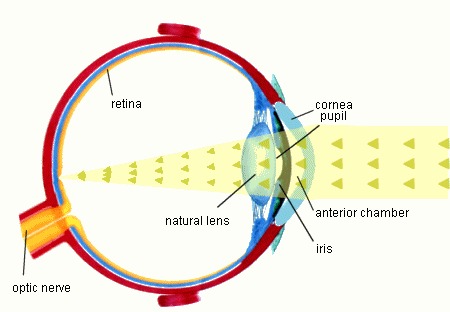
To understand how laser vision correction works, it helps to first understand the eye's natural optics and anatomical structures. In many ways, the eye works like an advanced camera: it focuses light rays from the outside world onto a light-sensitive "sensor" at the back of the eye — the retina. To see sharply, the eye's optical components must bend light rays so that they converge precisely on the fovea, the central area of the retina responsible for sharp, detailed vision.
The Cornea's Structure, in Brief
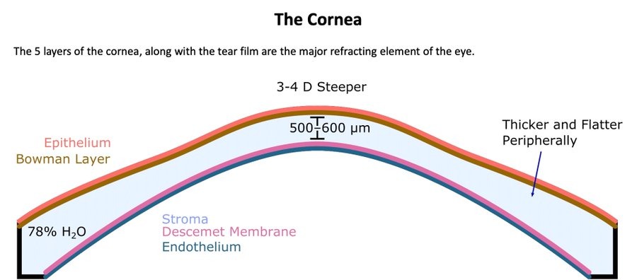
The cornea is the transparent "window" at the front of the eye, and it provides most of the eye's focusing power. It is made up of several thin layers: the outer layer (the epithelium) regenerates quickly if scratched or injured, while the thick middle layer — the stroma — is the layer that laser surgery acts on to permanently reshape the cornea and correct vision.
2. How Vision Works: Refractive Errors in Detail
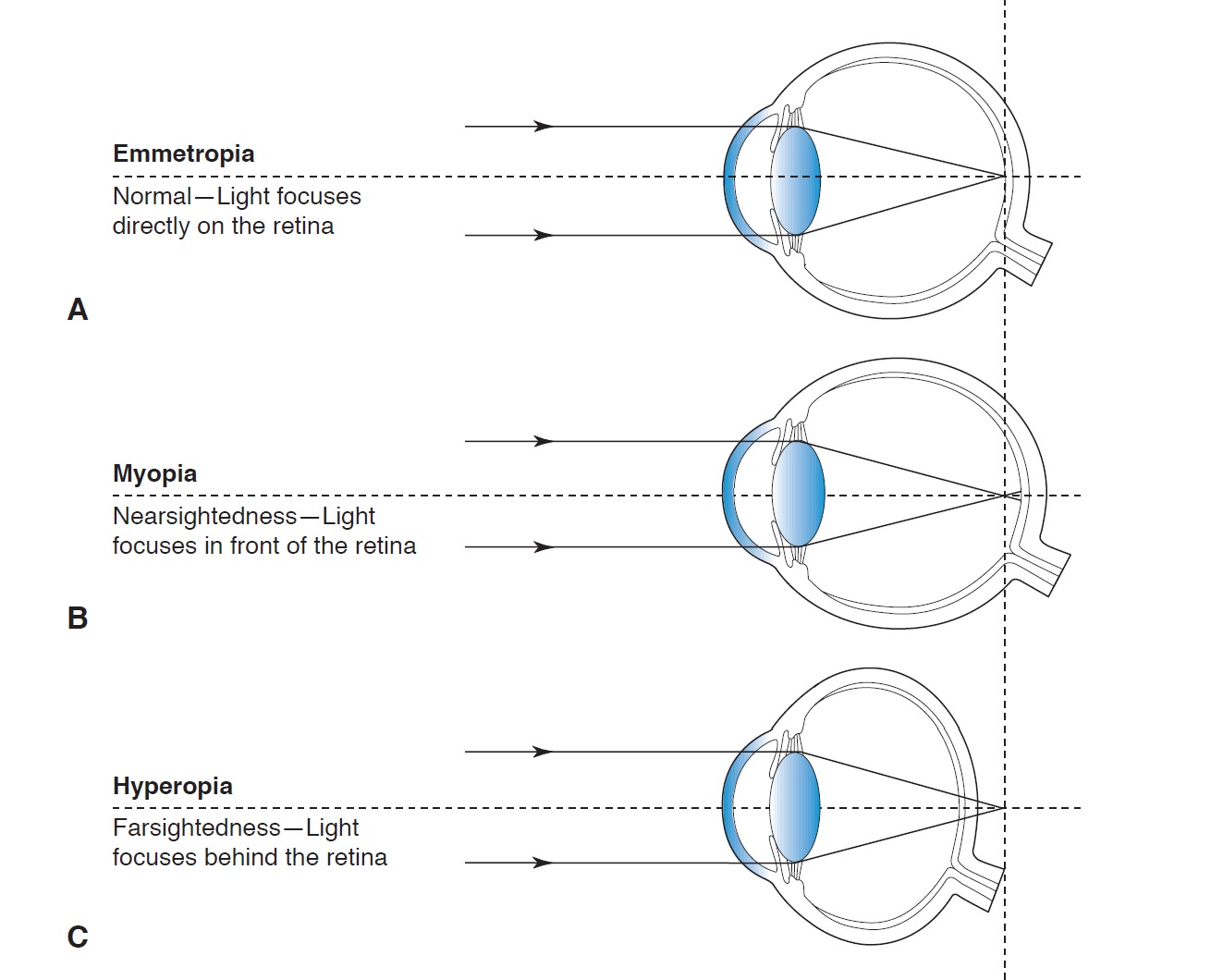
When the optical power of the cornea and natural lens does not perfectly match the physical length of the eye, light rays do not focus directly on the retina. This is called ametropia, or a refractive error. The goal of corneal refractive surgery is to reshape the cornea and its curvature so that the eye's focal point falls precisely on the retina.
Myopia
In a nearsighted eye, light rays from a distant object focus in front of the retina rather than on it — usually because the eye is too long or the cornea too steep. People with myopia see nearby objects relatively clearly, while distant objects appear blurred. Laser surgery corrects this by flattening the central cornea, which reduces the eye's focusing power and brings the focal point back onto the retina.
Hyperopia
In a farsighted eye, the opposite happens: light rays focus behind the retina, usually because the eye is too short or the cornea too flat. Distance vision is usually relatively good, but near vision requires constant focusing effort, which can cause eye strain and headaches. Laser surgery corrects this by adding curvature to the central cornea.
Astigmatism
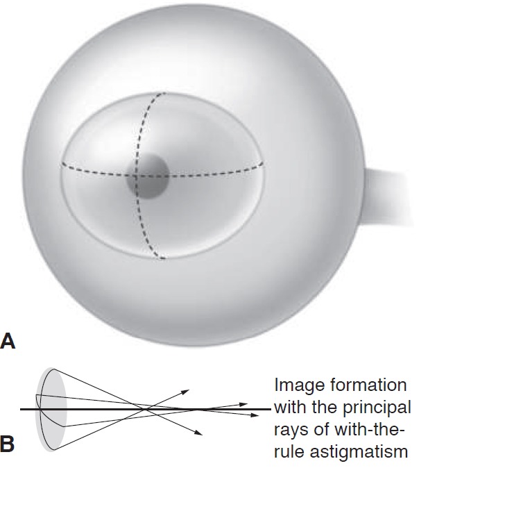
Astigmatism occurs when the cornea is not perfectly spherical but has different curvatures in different meridians. As a result, light focuses at two different points instead of one, and vision is blurred or distorted at all distances. Laser surgery corrects this through differential flattening — stronger treatment along the steeper direction — to create a more uniform surface.
Presbyopia — Age-Related Loss of Near Focus
Presbyopia is a natural, gradual loss of the ability to focus up close, usually starting around age 40, as the natural lens gradually loses its flexibility, making it harder to focus on near tasks. Because this is an aging process of the natural lens and not a problem with the cornea, laser corneal surgery does not "cure" presbyopia. It can, however, be planned for in advance using a monovision strategy.
Monovision for Presbyopia
In most cases, both eyes are targeted for clear distance vision. With monovision, by contrast, the dominant eye is set for distance vision and the other eye is set for mild nearsightedness, so that it provides better near vision. The brain learns to combine the two images. The difference between the eyes is set according to the patient's needs: a smaller difference better preserves depth perception, while a larger difference provides better near vision but may slightly reduce depth perception and cause halos at night.
3. Pre-Surgical Evaluation: Suitability and Safety
Not every eye is suitable for laser vision correction. Before recommending surgery, I perform a comprehensive evaluation to determine whether the procedure can be performed safely.
Prescription Stability
Before any laser procedure, it must be confirmed that the prescription has not changed significantly over the past year. Operating on an eye whose prescription is still changing can lead to a partial return of the refractive error after surgery, as the eye continues to change naturally.
Stopping Contact Lenses Before Your Evaluation
Contact lenses sit directly on the cornea and can temporarily distort its shape. To get accurate measurements before surgery, soft contact lenses must be stopped at least 1–2 weeks in advance, and rigid (RGP) lenses at least 3–4 weeks in advance.
Corneal Mapping
I perform detailed imaging of the shape and thickness of the cornea, to confirm it is healthy and stable enough for surgery, and above all to rule out keratoconus — a disease in which the cornea is thin and irregular from a young age, and in which laser surgery could cause harm rather than help.
When Surgery Is Not Performed
There are situations in which laser surgery is completely contraindicated: keratoconus, active eye infection or inflammation, uncontrolled glaucoma, severe untreated dry eye, and pregnancy or breastfeeding (due to temporary corneal changes). In other situations — such as poorly controlled diabetes, autoimmune diseases, or immune suppression — greater caution is required; both procedures call for individual judgement, and the choice between them is made case by case.
4. Laser Technologies
Laser vision correction surgery uses two complementary types of laser:
- Excimer laser: the laser that reshapes the cornea. It removes microscopic amounts of corneal tissue through precise photoablation, with minimal thermal effect on the surrounding tissue.
- Femtosecond laser: in these procedures it is used only in LASIK, to create the corneal flap gently and precisely, in place of a mechanical blade.
The treatment plan is personalized based on precise corneal mapping, and sometimes also on measurement of the eye's wavefront aberrations, to achieve the sharpest possible result and reduce effects such as glare and halos at night.
5. Modern Surgical Methods: PRK and LASIK — Step by Step
There are currently two main techniques for corneal laser vision correction: Photorefractive Keratectomy (PRK) and Laser-Assisted In Situ Keratomileusis (LASIK).
1. PRK — Surface Ablation
In PRK, the laser acts directly on the surface of the cornea, without creating a flap. First, the outer epithelial layer is gently removed, and then the excimer laser reshapes the exposed corneal surface. Finally, a soft therapeutic contact lens is placed, protecting the surface while the epithelium heals — usually within 3–5 days.
2. LASIK
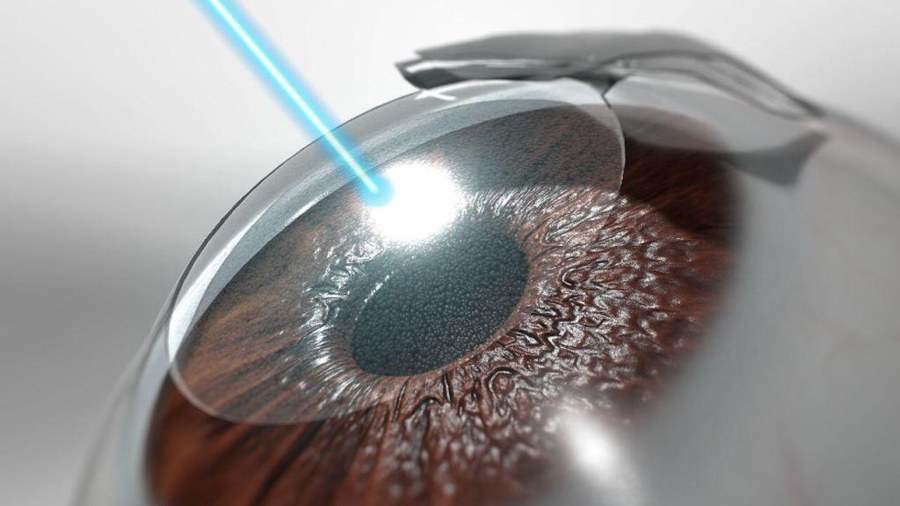
In LASIK, the femtosecond laser first creates a thin, uniform corneal flap, attached by a small hinge at its edge. I gently lift the flap and fold it back to expose the tissue beneath.
The excimer laser is then applied to the exposed tissue to reshape its curvature. At the end of the procedure, I reposition the flap and smooth it to ensure it lies flat without wrinkles. The flap adheres naturally within minutes, with no need for stitches.
6. Choosing the Right Procedure: A Comparison
To help patients understand the key differences, here is a comparison between the two main corneal refractive procedures across several clinical parameters:
| Parameter | PRK (Surface) | LASIK (Flap) |
|---|---|---|
| Typical range in practice* | Myopia: up to about −8.00 D Hyperopia: up to about +4.00 D Astigmatism: up to about 4.00 D |
Myopia: up to about −10.00 D Hyperopia: up to about +4.00 D Astigmatism: up to about 5.00 D |
| Flap-related risks | None — no flap is created | Rare risks of flap displacement, wrinkles (striae) or epithelial ingrowth at the interface |
| Discomfort | Moderate to severe for the first 3–4 days, until the epithelium closes | Minimal. Mild gritty or sandy sensation for 4–6 hours |
| Visual recovery | Slow. Functional vision within about a week; full stabilization over 3–6 weeks | Very fast. Most patients reach 20/20 as early as the first day |
| Dry-eye risk | Low. Minimal damage to the anterior corneal nerves | Moderate. Cutting the nerves temporarily reduces the tearing reflex |
| Biomechanical preservation | Greater. The strong anterior stromal lamellae are preserved | Lower than PRK. The flap interface remains a point of relative structural weakness |
| Lifestyle | Especially suitable for contact sports, military or police service; no flap that could shift | Contact sports or water activities should be avoided for 2–4 weeks |
* The approved ranges of the laser platforms are wider; the values in the table reflect the range within which most surgeons choose to operate, and in every case the decision depends on corneal thickness and shape.
7. Possible Risks After Surgery
Modern laser surgery is very safe, with satisfaction rates of over 90% in large clinical studies — but as with any surgery, it is important to be aware of the possible risks.
Undercorrection or Residual Refractive Error
The cornea is living tissue that heals somewhat differently in each patient, so a slight residual refractive error sometimes remains. In most cases this can be corrected with light glasses for demanding tasks such as night driving, or with a supplemental laser treatment once the prescription has stabilized.
Dry Eye
Nearly all patients experience temporary dry eye after surgery, due to temporary damage to the corneal nerves. In most cases this resolves completely within 3–6 months and is managed with artificial tears; in very rare cases, more prolonged discomfort can develop that requires dedicated treatment.
Corneal Ectasia
The most significant complication, but also the rarest: a structural weakening of the cornea that causes it to gradually bulge and become distorted, impairing vision. The risk of this is carefully assessed in advance — corneal thickness, the amount of tissue that will remain after surgery, and its shape — and can be substantially reduced through careful preoperative screening and candidate selection.
Other Rare Complications
With LASIK, there is a rare risk of a flap-related problem (displacement or mild wrinkles, mainly if the eye is struck in the first week) or mild inflammation beneath the flap, which is usually treated successfully with drops. These complications are significantly rarer in PRK, where there is no flap at all.
Risk of Retinal Detachment in High Myopia
Laser surgery only changes the shape of the cornea and does not change the physical length of the eyeball. Eyes with high myopia therefore remain at a lifelong increased risk of retinal tears and detachment, regardless of the surgery.
8. Frequently Asked Questions for Refractive Surgery Patients
How long does the procedure take, and is it painful?
The whole procedure takes about 10–15 minutes for both eyes, while the laser itself is active for only about 20–40 seconds per eye. The procedure is almost painless, since topical anesthetic drops are applied before surgery to numb the surface of the eye. During LASIK, you may feel a slight pressure sensation around the eye for a few seconds when the suction ring is applied.
How long is recovery, and when can I return to work?
After LASIK, recovery is very fast. Most patients achieve functional vision, often reaching 20/20, and return to work and normal computer use as early as the next day. After PRK, recovery is slower: expect moderate discomfort and blurred vision for the first 3–5 days while the epithelium heals, with vision gradually stabilizing over 3–6 weeks.
Can both eyes be treated on the same day?
Yes. Simultaneous bilateral treatment is standard practice in laser refractive corneal surgery. Correcting both eyes in the same session prevents anisometropia — a potentially very confusing condition in which one eye is fully corrected while the other is still significantly nearsighted or farsighted. That said, patients can choose a staged, one-eye-at-a-time approach if they prefer to first evaluate the visual result in the first eye before treating the second.
Will dry eye get worse after laser surgery?
Corneal laser surgery temporarily worsens dry-eye symptoms in almost all patients, due to temporary damage to the corneal nerves. In the vast majority of cases, symptoms can be controlled with preservative-free artificial tears, and they resolve completely within 3–6 months. Patients with moderate to severe dry eye before surgery should undergo ocular surface optimization before any laser procedure, using medications such as cyclosporine drops or punctal plugs.
Is laser vision correction permanent, or will I need surgery again in the future?
The structural change to the corneal stroma is permanent. However, the eye continues to undergo natural age-related changes. For example, laser corneal surgery does not prevent the natural development of presbyopia in your 40s; over time, reading glasses may therefore be needed for close-up work, unless a monovision correction strategy was chosen.
What happens if I blink or move my eye during the laser treatment?
There is no need to worry about blinking, because during the procedure a small, gentle lid speculum safely holds the eyelids open. To handle small eye movements, modern excimer systems are equipped with active, fast infrared eye-trackers. The trackers monitor the pupil hundreds of times per second; if the eye moves slightly, the laser immediately adjusts its target point. In the event of a sudden, large movement, the laser stops automatically and immediately, and resumes safely once the eye is repositioned.